B-mode of the ultrasound Download Scientific Diagram
4.6 (200) In stock


B-mode ultrasound images taken throughout the histotripsy process. (a)
How It Works: Ultrasound Modes
Breast B-mode ultrasound image conversion diagram; A, Regions of

Mechanical Wave Velocities in Left Ventricular Walls in Healthy Subjects and Patients With Aortic Stenosis - ScienceDirect
Examples for B-Mode ultrasound steatosis grading 0˚(0˚(A), I˚(I˚(B)

Ultrasound localization microscopy - ScienceDirect

B-Mode and PA-Mode imaging of different groups before and after

A Diagram of region of interest shown in B, C. B Standard high

B-mode ultrasound imaging measurement and 3D reconstruction of submerged topography in sediment-laden flow - ScienceDirect
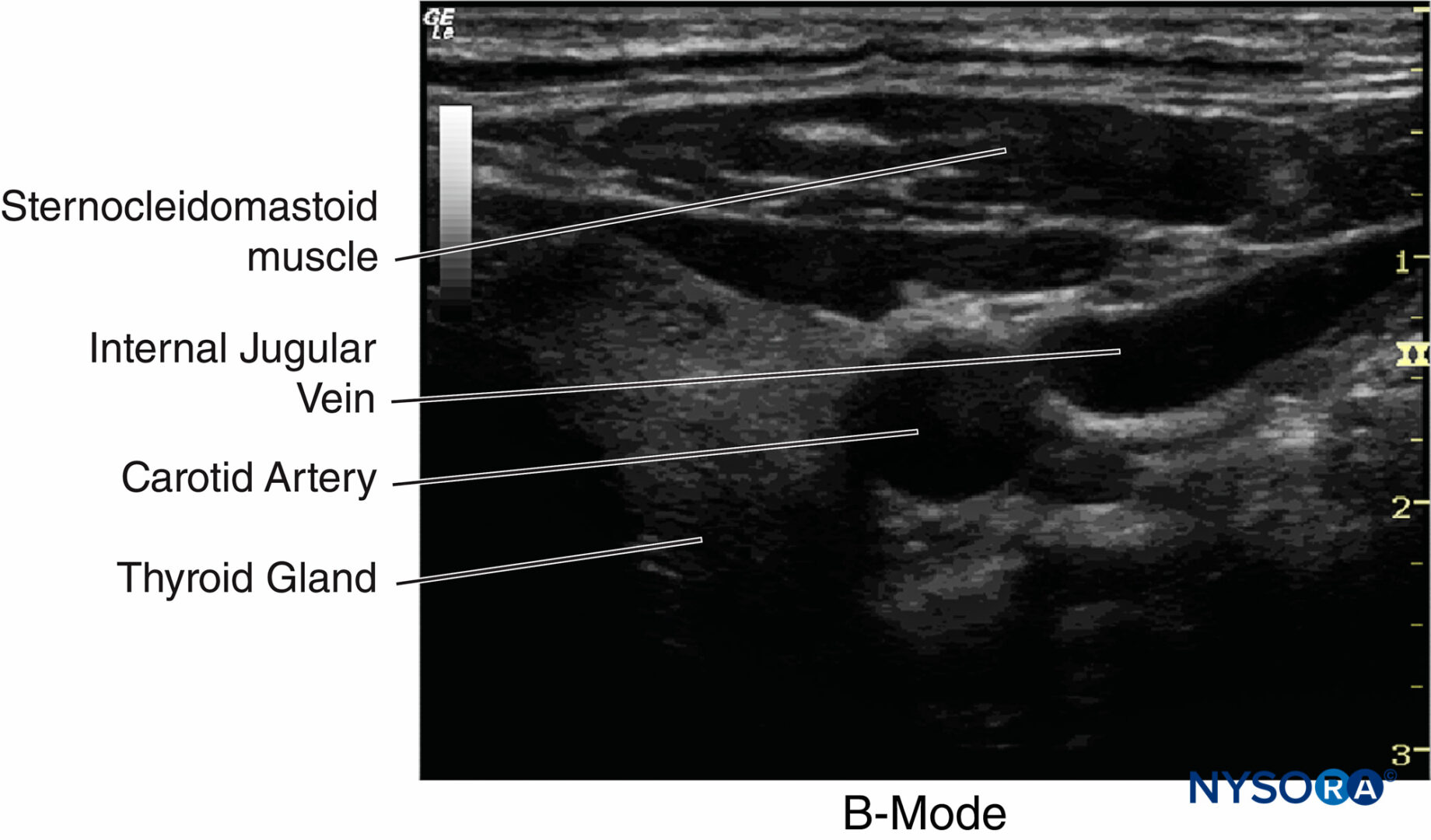
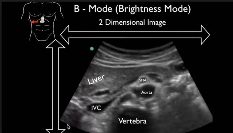
Physics of Ultrasound - NYSORA

B-mode ultrasound of left eye

Ultrasound Modes: A, B, & M Ultrasound, Sonography, Echo
In vivo time-harmonic ultrasound elastography of the human brain detects acute cerebral stiffness changes induced by intracranial pressure variations

A) B-mode ultrasound with MR fusion guidance shows a solid homogeneous
SonoZone: Ultrasound Modes: A, B, & M
B-Flow, Radiology Reference Article
Ultrasound imaging in B-mode, color and spectral Doppler of the
 20 gay swimwear brands you need to shop for this summer! • Nomadic Boys
 MRULIC yoga pants Twisting High Sports Tighter Camouflage Shorts
 Fashion sexy temptation bra ultra-thin full transparent gauze lace decoration small bra push up underwear female set - AliExpress
 ED HARDY by Christian Audigier For Women. Eau De
 Buy Lovable White Maternity Bra Non-Wired - 34B at
 Thinking Of You Dress - Black