Finite element analysis of compression fractures at the
5 (293) In stock
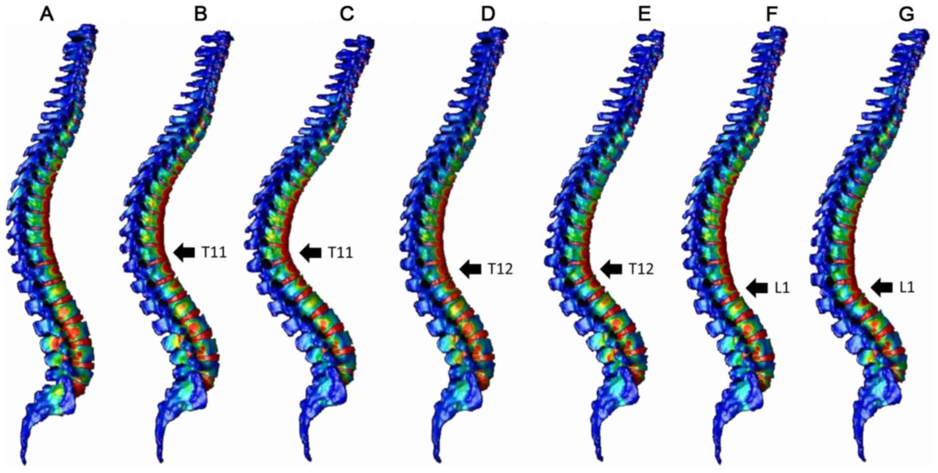
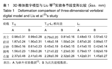
Vertebral fractures commonly occur at the thoracolumbar junction. These fractures can be treated with mild residual deformity in many cases, but are reportedly associated with increased risk of secondary vertebral fractures. In the present study, a three‑dimensional (3D) whole spine model was constructed using the finite element method to explore the mechanism of development of compression fractures. The 3D model of the whole spine, from the cervical spine to the pelvis, was constructed from computed tomography (CT) images of an adult male. Using a normal spine model and spine models with compression fractures at the T11, T12 or L1 vertebrae, the distribution of strain was analyzed in the vertebrae after load application. The normal spine model demonstrated greater strain around the thoracolumbar junction and the middle thoracic spine, while the compression fracture models indicated focused strain at the fracture site and adjacent vertebrae. Increased load time resulted in the extension of the strain region up to the middle thoracic spine. The present findings, that secondary vertebral fractures commonly occur around the fracture site, and may also affect the thoracic vertebrae, are consistent with previous clinical and experimental results. These results suggest that follow‑up examinations of compression fractures at the thoracolumbar junction should include the thoracic spine and adjacent vertebrae. The current data also demonstrate that models created from CT images can be used for various analyses.

Finite Element Method Analysis of Compression Fractures on Whole-Spine Models Including the Rib Cage
Treatment of cervical vertebral (C1) metastasis of lung cancer with radiotherapy: A case report

Finite element model analysis of bidirectional compression porous
Comparative analysis of tibial plateau fracture osteosynthesis: A finite element study,Journal of the Mechanical Behavior of Biomedical Materials - X-MOL
Biomechanical analysis of four augmented fixations of plate osteosynthesis for comminuted mid‑shaft clavicle fracture: A finite element approach
Finite Element Method Analysis of Compression Fractures on Whole-Spine Models Including the Rib Cage - Document - Gale OneFile: Health and Medicine

Biomechanical investigation of long spinal fusion models using three-dimensional finite element analysis, BMC Musculoskeletal Disorders
Mechanical changes of percutaneous kyphoplasty and percutaneous vertebroplasty in the treatment of thoracolumbar compressive fractures in three-dimensional vertebral models
Finite element analysis of tibial fractures

Finite element analysis of a novel anatomical locking plate for scapular neck fracture, Journal of Orthopaedic Surgery and Research

PDF) Optimal Fixation of Acute Scaphoid Fractures: Finite Element Analysis

Biomechanical investigation of long spinal fusion models using three-dimensional finite element analysis, BMC Musculoskeletal Disorders
Frontiers Biomechanical Evaluation and the Assisted 3D Printed Model in the Patient-Specific Preoperative Planning for Thoracic Spinal Tuberculosis: A Finite Element Analysis

Bioengineering, Free Full-Text
Cureus, Complex Compression Fracture in the Thoracolumbar Junction: A Case Report
Preoperative MRI and X-ray show a L3 compression fracture with
Compression Fracture Treatment
Compression Fracture of the Spine - Plainsboro Township, NJ
Vertebral Compression Fracture Vancouver - Dr. Craig Best - Dr. Craig Best
 Always Infinity Super Unscented Pads w/Wings Flex Foam Size 2 - 16ct/1 – My Store
 My Honest Excellence Playa Mujeres Review - The Daley Dose
 Beige Linen Long Sleeve Shirt Outfits For Men (37 ideas & outfits
 Tiny Cup Trophy Crochet pattern by Lottie's Creations, Knitting Patterns
 Cupid's Undie Run Is the Hottest Race in St. Louis [PHOTOS]
 Concord Edwardian era Fashion Suit Male Edwardian fashion men, Edwardian era fashion, Vintage mens fashion