Medial view of left knee region highlighting various fascial
4.7 (483) In stock

Download scientific diagram | Medial view of left knee region highlighting various fascial components surrounding the semitendinosus muscle. From the superficial to the deep aspect: the fascia lata, the paratenon and the epimysium from publication: Anatomical study of paratenons and fascia lata connections in the posteromedial knee region | Introduction In the last decade, fascia research increased significantly in various aspects such as anatomical and biomechanical features related to epimuscular force transmission. Methods The present anatomic study focuses on macroscopic observations of the potential | Fascia Lata, Hamstring muscles and Fascia | ResearchGate, the professional network for scientists.

Medial view of left knee region highlighting various fascial

Medical Stock Image - Healthy Knee Labelled Anterior View

Key Surgically Relevant Anatomy of the Medial and Lateral Aspects

Key Surgically Relevant Anatomy of the Medial and Lateral Aspects
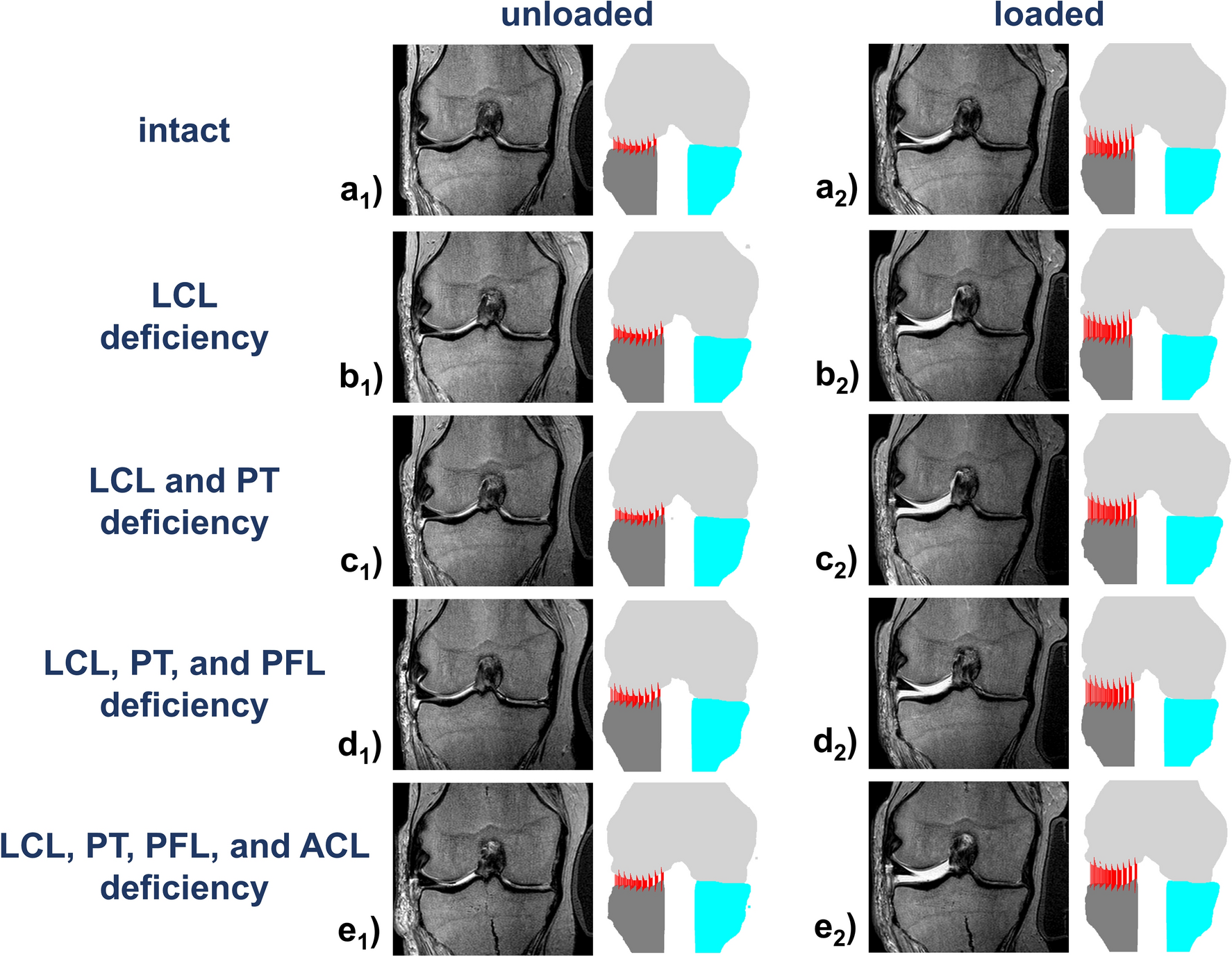
Varus stress MRI in the refined assessment of the posterolateral

Medicina, Free Full-Text

Key Surgically Relevant Anatomy of the Medial and Lateral Aspects
Calf Strain - Physiopedia

Tensor Fasciae Latae (TFL) Muscle Anatomy - Bodyworks Prime
/images/vimeo_thumbnails/258296721/UWQ0ucbOB1jHt8YDVJ8bQQ_overlay.jpg)
Perineal region: Anatomy, definition, diagram
The tensor fascia lata (TFL) is - NeuroKinetic Therapy
Fascia Lata and Iliotibial Tract Diagram
Tensor fascia lata muscle, illustration - Stock Image - F026/9468 - Science Photo Library