Coronal and axial slices displaying the IFG area that showed
4.6 (110) In stock

Changes in brain structure and function in a multisport cohort of retired female and male athletes, many years after suffering a concussion. The ICHIRF-BRAIN Study

2 Anatomy and Development

Coronal, sagittal and axial slices of the brain depicting the regions

Hypothalamus and amygdala functional connectivity at rest in narcolepsy type 1. - Abstract - Europe PMC

Matthew C Hagen's research works University of Minnesota Duluth

David ZALD, Professor (Full), Ph.D.

Human Brain Mapping, Neuroimaging Journal

José PARDO Professor (Full); Director, Cognitive Neuroimaging
Axial, Coronal & Sagittal Planes
Tricia THORNTON-WELLS, PhD, Neuroscience

Stroke progression, Radiology Case
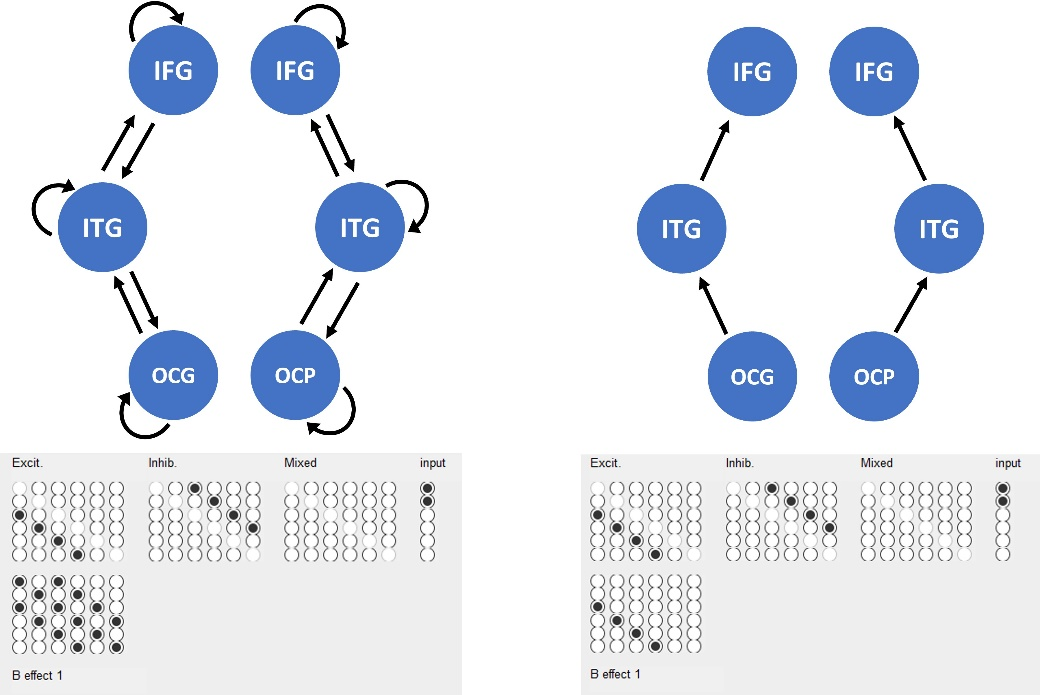
DCM for Evoked Responses - SPM Documentation

Frontiers Functional Connectivity of the Human Paraventricular Thalamic Nucleus: Insights From High Field Functional MRI

Retrieving meaning after temporal lobe infarction: The role of the basal language area - Sharp - 2004 - Annals of Neurology - Wiley Online Library
How IFG and Google Cloud AI bring structure to unstructured financial documents
We want to equip government to deliver better': IfG director sets
Visual word processing engages a hierarchical, distributed, and