B-mode ultrasound, color Doppler, and sonoelastography in
4.6 (266) In stock
Background Enlarged cervical lymph nodes are the most commonly encountered neck lumps. Ultrasonography is the most extensively used tool for classification of superficial lymph nodes due to its availability and low cost. Ultrasound (US) elastography refers to a non-invasive imaging technique that can describe tissue displacement (i.e., strain) or stiffness in response to a given force. The aim of this study is to compare between B-mode sonography, color Doppler, and sonoelastography in assessment of enlarged deep cervical lymph nodes. Results The prevalence of benign lymph nodes was 26 out of 84 (31%). Lymphomatous lymph nodes were 22/84 (26.2%), while metastatic lymph nodes were 36/84 (42.8%). Color Doppler evaluation of nodal vascular pattern was of high sensitivity (91.7%), specificity (80.8%), and accuracy (88.6%) for differentiating metastatic and benign nodes (P value was < 0.001). There was a significant difference between elasticity scores of benign and malignant lymph nodes (P < 0.001). The most frequent score in the malignant group was 3 (21/27) (77.8%) while the most frequent score in the benign group was 2 (5/11) (45.5%). The mean strain ratio (strain index) for malignant lymph nodes (mean 3.2 ± 0.8) was significantly greater than that for benign lymph nodes (mean 1.1 ± 0.8). Conclusion Ultrasound elastography with its high sensitivity and specificity is a helpful improvement in US for the assessment of cervical lymph nodes, in which biopsies should be performed.

Cystic Lymph Node Metastases in Papillary Thyroid Carcinoma

Egyptian Journal of Radiology and Nuclear Medicine
Cureus Role of Sonoelastography in Differentiating Benign From

Utility of Sonoelastography Beyond Sonography for Differentiation

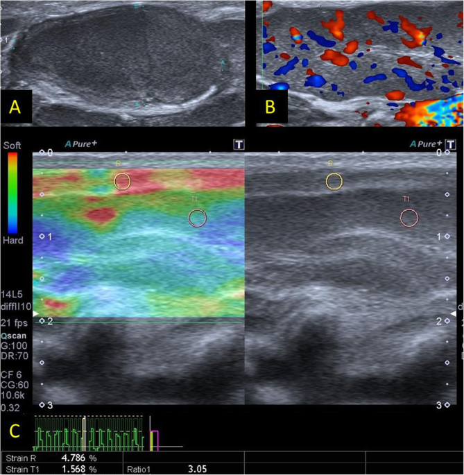
US elastogram shows blue areas almost entirely filling the lymph

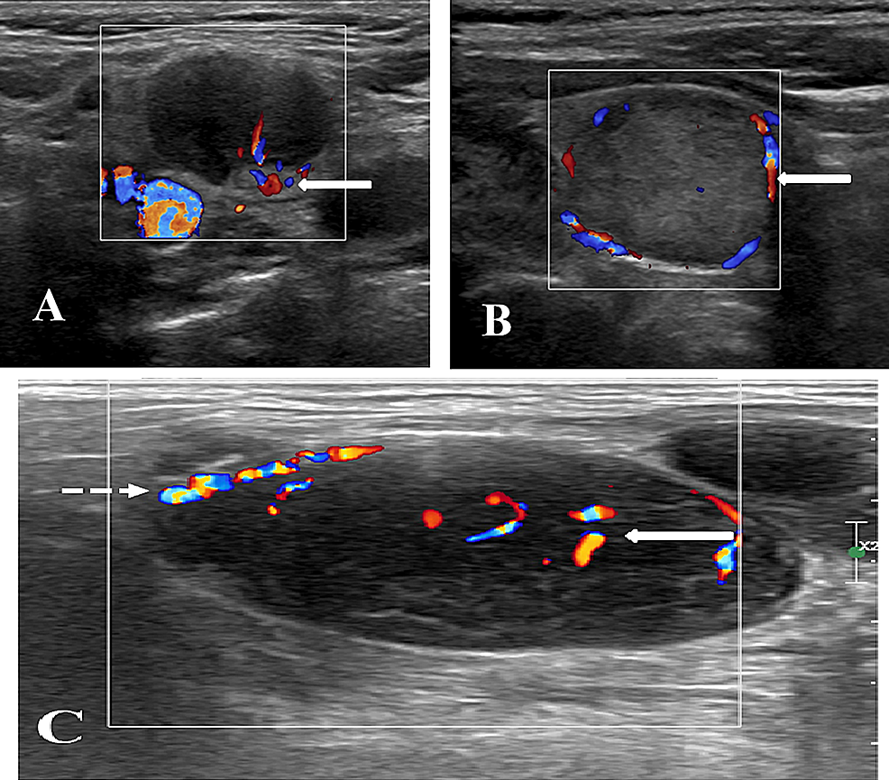
B-mode US (a) revealed a well-defined hypoechoic left lower deep

Reliability of sonoelastography in predicting pediatric cervical

Application of multimodal ultrasonography for differentiating

Ehab ABDELGAWAD, Minia University, Al Minyā

Application of multimodal ultrasonography for differentiating

a and b) Split-screen B-mode ultrasound image (left) and US

Ehab ABDELGAWAD, Minia University, Al Minyā

PDF) B-mode ultrasound, color Doppler, and sonoelastography in

Utility of Sonoelastography Beyond Sonography for Differentiation
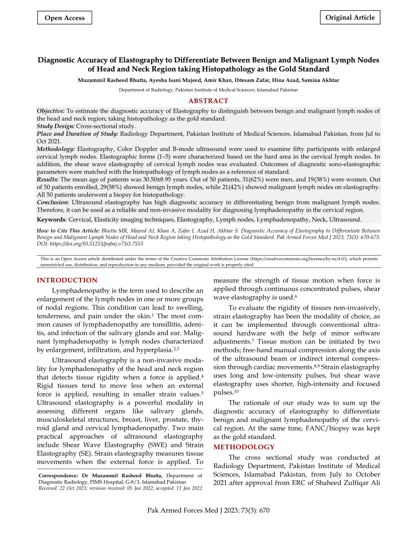
PDF) Diagnostic Accuracy of Elastography
Ultrasound images • Liver, B-mode, echogramm №847
A-mode and B-mode ultrasound measurement of fat thickness: a cadaver validation study
Pitfalls of inferior vena cava M-mode – NephroPOCUS
Prius Shifter B Mode: Everything You Need To Know
Ultrasound imaging in B-mode, color and spectral Doppler of the
 Yingfa9117 long swimming pants legskin swim trousers swim jammers for men & boys
 Nike Women's Pro Classic Swoosh Medium Support Sports Bra Heather Gray XS
 Plus size see through lingerie female
 28/30/32/34 Inseam Women's Bootcut Yoga Pants Long Bootleg High-Waisted Flare Pants with Pockets
 Under Armour Sneakers for Men, Online Sale up to 49% off
 Zip Compression Socks Medical, 2 Pair Toeless Nurse Compression Socks with Zipper Easy on off 15-20 mmHg for Varicose Veins, Edema, Swollen or Sore